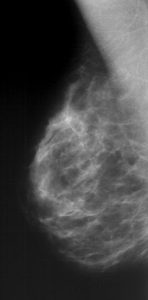
- Mammography, mammogram
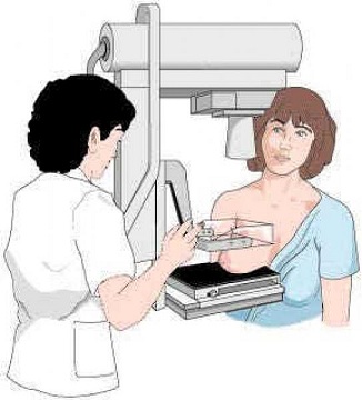
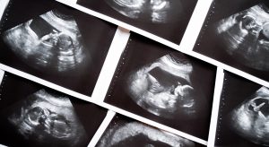
It is an X-ray examination of the breast to detect the beginning of a cancerous growth. It is the basic imaging method for most women. As a rule, it is not suitable for young women in whom the gland is rich and dense. Mammography is performed on a machine, the mammograph, which uses so-called soft X-rays. During the examination, the breast must be sufficiently compressed, this is necessary to achieve a high quality image. Usually the breast is examined in two planes, so a total of 4 images are taken.
Some mammography examinations must be supplemented by ultrasound. In our department, this examination is performed immediately on the same day, within one patient visit. For scheduled examinations, it is optimal to examine in the first half of the menstrual cycle, when the breasts can be sufficiently squeezed without discomfort for the woman being examined, and when there is a risk of delay (if a malignant tumour is suspected).What is mammography screening?
Mammography screening is a regular preventive examination of women without any signs of disease in order to catch developing disease at the earliest possible stage. The principle of mammography screening is based on the premise that disease caught at an early stage is more easily treatable and leads to a higher quality and longer life for patients.
Breast cancer is currently one of the most serious epidemiological problems in the Czech Republic. More than 4,600 new cases are diagnosed every year in our country and the incidence is constantly increasing. More than 2 000 Czech women die of breast cancer every year and breast cancer is the leading cause of death in the age group 20-54 years.
There are limited options for disease prevention at the individual level. Since the risk factor explaining the cause of this disease has not yet been identified with satisfactory reliability (as is the case with lung cancer), early diagnosis and successful treatment are the only options for its control.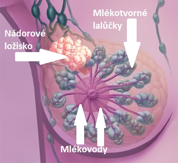
Examination
Diagnostic mammography - is performed in patients with symptoms of breast cancer or already diagnosed breast cancer or as a complementary examination to screening mammography.
Screening mammography - is performed in accordance with the relevant decree of the Ministry of Health of the Czech Republic within the framework of preventive examinations and dispensary care. It is not an appropriate method for women under 40 years of age (except for women with a significantly increased risk of breast cancer, who are monitored according to an individually determined scheme) and should not be performed more frequently than annually. A woman should come to the screening mammography centre with a request to undergo this procedure or with an invitation (age-based indication). A female patient may come directly to the screening mammography centre without a request.Ductography - is indicated in patients with spontaneous pathological secretion from the nipple, which could be a manifestation of cancer (papilloma, carcinoma, etc.).
Preparation:
- Examination performed standing, it is necessary to remove all cosmetic products in the armpits.
- Ultrasound examination - without preparation, examination lying down.
- Introduction of the locator to non-palpable bearings - without preparation, in cooperation with the requesting department.
- Percutaneous puncture or ultrasound-guided biopsy - without preparation, examination lying down, possible risks - allergic reaction to numbing agent or risk of bleeding and haematoma. Restrictions after the procedure - cover dressing until the next day and restriction of physical work and sports activity on the day of the procedure.
Examinations covered by the public health insurance
The screening is covered by the public health insurance once every two years for every woman over the age of 45. However, to avoid having to pay for the screening, you must bring a valid application form to the surgery. A request form is actually a request written on paper in which your doctor asks you to have a preventive breast examination. What does this mean? To have a preventive breast examination, you must first visit your gynaecologist or GP, who is obliged to issue you with a request form. The referring doctor then has the right to know the result of the examination and you are obliged to give this report to him. This procedure may seem unnecessarily complicated, but it is not. It is imperative that the doctor whom the woman sees the most knows whether his client is healthy and can safely prescribe all the medicines she needs. A woman who has a breast tumour, for example, cannot take hormonal drugs to the same extent as a healthy woman, and her doctor must know this fact.
The examination is paid for by you
Women who pay for their own mammograms or ultrasounds (self-payers) can be examined without a request (voucher) from their doctor. This applies to: Women who pay for their own mammograms or ultrasounds (self-payers) can be examined without a doctor's order (examination voucher). This applies to:
1) Women in the screening population (over 45 years of age) who wish to be screened earlier than the screening interval (currently 2 years between two consecutive mammograms),
2) women outside the screening population - i.e. those under the age of 45.Appointment / enquiry about a preventive (screening) examination can be made by calling 567 157 602,
or through the online form - HERE
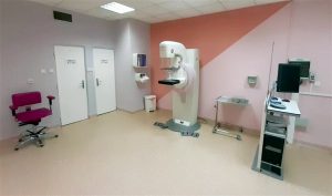
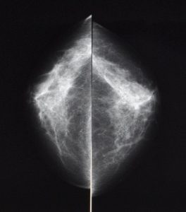
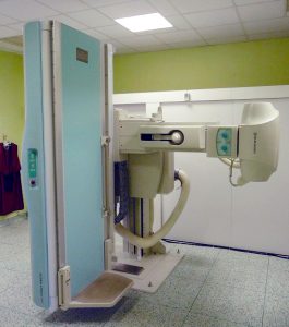
Instrumentation and sample images:
- Mammomat Inspiration, Siemens
- Sciascopy
Sciascopy
It is a radiological examination method that uses X-rays to view the human body in real time. It is mainly used for imaging of the digestive tube (esophagus, stomach, duodenum, small and large intestine), imaging of the biliary tree (ERCP), less often for imaging of other hollow organs (bladder, uterine cavity), spinal canal or some pathologies (fistulas...), etc.
Contraindications to the examination
- Pregnancy.
- An uncooperative patient.
Particular examinations:
X-ray examination of the esophagus - standing up, no preparation.
X-ray examination of the stomach and duodenum, small intestine, passage through the digestive tube - the examination is performed standing, the patient must not eat, drink or smoke from midnight on the day of the examination. The patient should come for the examination in the morning, when there is a minimum of gastric juices in the stomach. In patients whose pylorus is narrowed, the stomach contents must be drained.
X-ray examination of the colon -irrigography - the examination is performed lying down. Before the examination, the patient's bowel must be emptied. Emptying is carried out by adjusting the diet and infusions or with the help of medication. The diet should be porridge-like and fat-free for 2-3 days before the examination. The patient presents for the examination with a rectoscopic finding - without it the examination cannot be performed.
X-ray examination - Perioperative or T-scan cholangiography - the examination is performed lying down in the surgery room.
X-ray examination of the urinary bladder -cystography - examination performed lying down, in cooperation with a urologist. To detect urinary retention in the bladder.
X-ray micturition cystourethrography - examination performed lying down. In collaboration with a urologist or the pediatric department. To determine the reflex in the ureters.
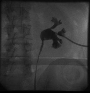
Retrograde single-sided pyelography - examination performed lying down. Imaging of the hollow renal system. Examination carried out in collaboration with a urologist.
Antegrade single-sided pyelography - examination performed lying down. Imaging of the hollow renal system by percutaneous puncture, examination performed in collaboration with a urologist.
X-ray examination of the uterine cavity and fallopian tubes - Hysterosalpingography - examination performed lying down, in cooperation with a gynaecologist. The patient arrives defecated, just before the examination she urinates, nervous women should be premedicated. Contraindications - acute inflammation, bleeding, pregnancy.
ERCP - endoscopic retrograde cholangiopancreatography - examination performed lying down. ERCP is a demanding, invasive examination of the biliary and pancreatic ducts, which is performed endoscopically under X-ray control. It is always performed during hospitalization. The patient is fasting from midnight, can take a sip of tea or water in the morning, and the p.o. medication is discontinued. Before the ERCP examination, the patient should not have an X-ray examination of the digestive tract.
You can make an appointment / inquire about an examination by calling 567 157 602
Instrumentation and sample images:
- Siemens Artis zee
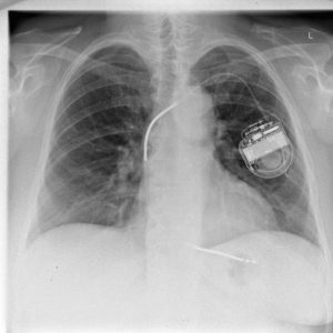
- Sciagraphy
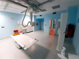
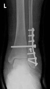
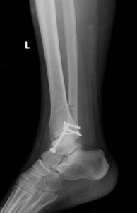
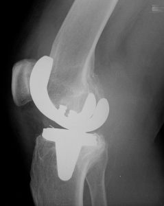
is a basic radiological examination performed in outpatient and inpatient facilities, indicated and acute. It is based on the principle of different absorption and scattering of X-rays in individual tissues of the human body in the direction of the focal point - image receiver. The resulting image then provides clear information about the structures under observation, which are the skeleton, soft tissues and structures with contrast agent filling. The scanning imaging system enables high contrast and high spatial resolution imaging with the possibility of achieving low radiation burden to the patient.
Contraindications to the examination
- Pregnancy.
- An uncooperative patient.
Examinations performed:
X-ray examination of the fingers and metacarpals of the hand or foot.
X-ray examination of the skull, targeted images.
X-ray examination of the skull, overview images.
X-ray examination of the neck and cervical spine.
X-ray examination of the thoracic and lumbar spine.
X-ray examination of the sacrum and SI joints.
X-ray examination of the pelvis or hip joint.
X-ray examination of the shoulder joint.
X-ray examination of the bones and hips of the extremities (excluding the brachial and pelvic plexus, fingers and metacarpals of the hand or foot).
X-ray examination of the ribs and sternum.
X-ray of the chest.
X-ray examination of the joint-held images.
X-ray examination of the limbs using the soft imaging technique.
X-ray of the abdomen.Instrumentation and sample images:
- Siemens Multix Fusion, Siemens Ysio Max a Siemens Ysio X.pree
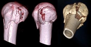
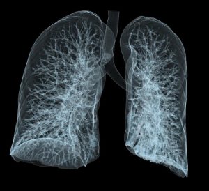
- CT scan - computed tomography
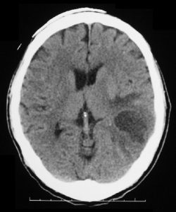
It is a device that works with X-rays in a similar way to a conventional X-ray machine, but in CT (computed tomography) the radiation source - the X-ray tube and the detection system - rotates around the patient's body, which is then irradiated gradually from different angles around the body. The detection system then evaluates the amount of radiation passing through the patient's body at different angles and from this data, individual scans - images of the body layers, usually in the axial plane - are then reconstructed using a powerful computer.
In a classical X-ray, the result is a summative image of the whole body in which the shadows of individual structures overlap; in a CT scan, we obtain thin layers of the examined area, without overlapping individual parts of the body, so it is possible to show individual organs, their structure and pathologies much more accurately.

Indications for examination
Due to the relatively high burden of X-rays, CT is indicated as a "second choice method" - a complementary examination that should help to clarify unclear findings of ultrasound or conventional X-rays, especially in the assessment of bones, lungs and intestines, where ultrasound is not applicable. For acute (urgent) indications, we use CT mainly for head-brain imaging in stroke and head trauma as it will show intracranial haemorrhage very well. Traumatic changes of the chest, abdomen, pelvis and bone fractures are also very reliable. Another very important acute indication for CT scanning is suspected aortic (heart) involvement, such as an aneurysm or dissection (tearing of the lining) of the aorta. In this case, CT angiography (CT examination of blood vessels) is performed, mainly to image the large arteries (aorta, renal arteries, pelvic arteries, carotid arteries) and also to image the cerebral arteries (circulus arteriosus Willis). In some cases, it can completely replace conventional angiographic examination.

Contraindications to the examination
Before the examination, it is necessary to make targeted inquiries and to exclude or confirm the presence of contraindications. Each patient is instructed by the attending physician about the risks of intravenous contrast agent administration and, if it is anticipated that it will be administered during the CT scan, the attending physician requests and countersigns the patient's written informed consent for its intravenous administration.
For relative contraindications, the risk-benefit ratio of the test must be considered.
Native examination
Gravity - only in serious danger to life.
Examination with a contrast agent
The examination must not be performed in patients allergic to iodine contrast agents.
In the cases listed below, a contrast CT scan may be performed only in emergency cases (vital indications - the scan must be performed to save the patient's life) and under emergency measures specific to the case (thorough and extended anti-allergic preparation, assistance of the anaesthesia team, provision of haemodialysis, necessary hospitalisation, etc.):
• pregnancy,
• hepatic and renal insufficiency, followed by dialysis,
• hyperthyroidism (increased thyroid function),
• pheochromocytoma (catecholamine-producing tumor),
• an uncooperative patient.Risks of examination
The CT machine uses X-rays and therefore there is a certain radiation load on the patient, see section "Ionizing radiation".
Administration of a contrast agent (intravenously or orally) - may cause adverse effects: hot flushes, sweating, nausea, redness, itching, rash and, in very rare cases, a severe allergic reaction in the form of anaphylactic shock may occur, requiring medical attention and, exceptionally, hospitalisation of the patient.
You can make an appointment / inquire about an examination by calling 567 157 561
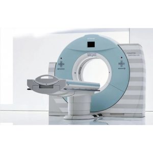
Instrumentation and sample images:
- CT Siemens SOMATOM DEFINITION AS+ 128CT The Siemens Somatom Definition AS+ allows simultaneous scanning of 128 layers per turn of the X-ray tube. The exceptional spatial resolution offers imaging of tissue structures as small as 0.24 mm, and the image scanning range of 200 cm combined with this resolution enables examination of the entire patient body in 10 seconds. This is particularly important when examining patients with severe trauma, unconscious or uncooperative patients. These exceptional parameters are particularly useful in traumatology, where it is important to make a diagnosis as quickly as possible, and in cardiology, where they allow examination of cardiac function and evaluation of coronary arteries, including analysis of calcifications in their walls.The speed and quality of the images are advantageous for qualitative vascular diagnosis and planning of interventional procedures on blood vessels. Fully automatic 3D volume imaging facilitates the evaluation of cerebral blood flow. Images from this device are generated in DICOM format, allowing immediate transmission to the PACS system and viewing of the submitted CT images at clinical sites.
- CT Siemens SOMATOM DEFINITION AS+ 128CT The Siemens Somatom Definition AS+ allows simultaneous scanning of 128 layers per turn of the X-ray tube. The exceptional spatial resolution offers imaging of tissue structures as small as 0.24 mm, and the image scanning range of 200 cm combined with this resolution enables examination of the entire patient body in 10 seconds. This is particularly important when examining patients with severe trauma, unconscious or uncooperative patients. These exceptional parameters are particularly useful in traumatology, where it is important to make a diagnosis as quickly as possible, and in cardiology, where they allow examination of cardiac function and evaluation of coronary arteries, including analysis of calcifications in their walls.The speed and quality of the images are advantageous for qualitative vascular diagnosis and planning of interventional procedures on blood vessels. Fully automatic 3D volume imaging facilitates the evaluation of cerebral blood flow. Images from this device are generated in DICOM format, allowing immediate transmission to the PACS system and viewing of the submitted CT images at clinical sites.
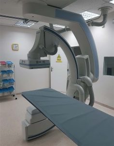
- Angiography and Interventional Radiology
What is Angiography?
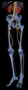
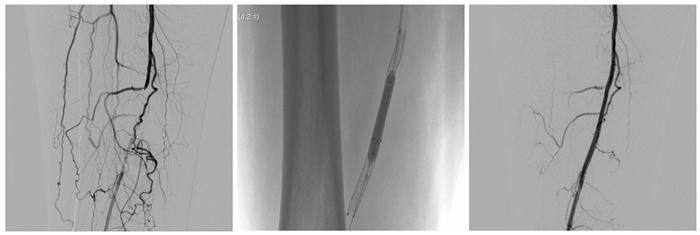
Angiography is an examination that is performed using X-rays and a contrast agent that is injected into a blood vessel. The examination is mainly used to view the arteries. The classic requirement is to examine the arterial system of the lower limbs when ischemic disease of the lower limbs is suspected. In addition, narrowing of the renal arteries or narrowing of the carotid arteries (neck arteries) can be detected. This method of examining the vascular supply is slowly being superseded by CT angiography and MR angiography in which it is not possible to perform a therapeutic intervention at the same time - angioplasty (stretching the vessel with a balloon) or the insertion of a reinforcement in the form of a metal tube (stent).
The process of examination
The examination is performed lying down under local anaesthesia. The patient lies on the examination table in the angiography room. The doctor numbs the puncture site, e.g. groin (possibly another entry site) and punctures the femoral artery (or another artery depending on the entry site). A thin plastic tube is inserted into the artery using a guide wire and a contrast medium is then passed through it. An X-ray machine is available at the examination table, on the screen of which the doctor can see in real time the flow of the contrast medium through the arteries and thus find pathologically affected areas on the vessels. The examination takes about one hour on average, including any therapeutic procedure. The X-ray images are stored in a computer and the doctor can also look at them at any later time.
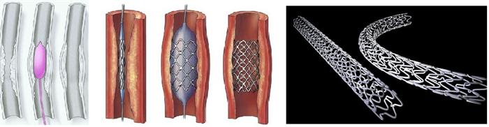
If a narrowed area is found, the doctor can dilate it with a tiny balloon that is inserted into the designated area through a thin wire (lead) in the blood vessel. In addition, a reinforcement in the form of a metal tube (stent) can be inserted to reinforce the narrowed area.
After the examination is completed, the puncture site is covered with a firm bandage (bandage) and a pressure bandage or the puncture site is closed with a special vascular patch, which is later absorbed by itself. The patient must lie still for 8 hours after the procedure and not get out of bed. The pressure dressing is usually removed after 8-12 hours.
Contraindications to the examination
As with other examinations where the patient is given a contrast agent, an allergic reaction may occur. The contrast agent also places a burden on the kidneys and can damage them. For this reason, a drinking regimen should be followed after angiography or intravenous infusions can be used.
Thyrotoxicosis - increased thyroid functionBenefits of the examination (procedure)
- A high-quality view of virtually all blood vessels in the human body.
- The possibility of direct therapeutic intervention for some arteries or veins in the human body with a simple puncture (minimally invasive procedure) without the need for surgery.Disadvantages and possible complications of the examination (procedure)
The device used for the angiographic examination works with X-ray ionizing radiation, which results in a radiation burden on the patient; therefore, it is necessary to indicate this examination with regard to the magnitude of this burden and the diagnostic and therapeutic yield.
A hematoma (bruise) may form at the puncture site, therefore it is necessary to observe bed rest for 6 to 24 hours, bleeding may also occur in the vicinity of the puncture, possibly even thrombosis of the vessel. These complications are managed conservatively or surgically depending on their extent and nature.
Potential complications:
- Disruption and release of the atherosclerotic plaque with subsequent injection into the vasculature.
- Rupture of aneurysm during surgery with bleeding.
Extravascular procedures
The extravascular interventional procedures performed in our department include mainly puncture and drainage of pathological fluid inflammatory collections in the abdominal cavity, thoracic cavity, retroperitoneum or pelvis. Diagnostic biopsies of pathological deposits in various organs.
You can make an appointment / inquire about an examination by calling 567 157 645
Instrumentation:
- Siemens Artis Zee Ceiling
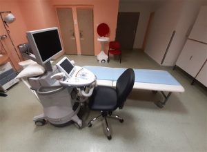
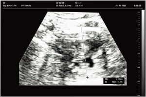
- Ultrasound
It is a painless, non-invasive examination that uses high-frequency sound waves (not ionizing X-rays) to image the tissues of the human body, and through which the doctor can make a diagnosis and suggest further treatment. Ultrasound waves travel through the body and reflect off individual organs or transitions between tissues with different acoustic impedances. The disadvantage is the inability of ultrasound to pass through bone and air, making it impossible to examine, for example, the lungs or loops of intestine. During the examination it is necessary to exert sufficient pressure on the measuring probe for good contact with the patient's body, which can be uncomfortable, therefore the surface of the probe is also covered with a gel (ECG cream) to avoid air gaps between the probe and the body of the examined. Because the tissues are imaged in real time during the ultrasound examination, it is possible to capture the structure and movement of the individual organs, as well as the blood flow in the blood vessels.
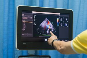
The Doppler effect, described in 1842, is used to measure blood flow. In a Doppler measurement ("colour Doppler"), the relative movement of the blood relative to the probe emitting the ultrasound is displayed by different coloured fields within the sections through the blood vessels or heart. It is therefore possible to detect any narrowing or blockage of blood vessels or, for example, an atrial defect in the heart.
Harmful effects of the examination?
No adverse side effects have been demonstrated during the time ultrasound has been used in medicine, but it should only be used for clear indications, on technically suitable equipment, and should always be performed by a qualified physician.
Indications for an ultrasound examination
The examination is performed to assess the morphological findings in the area of interest. In particular, it is an assessment of the organs in the abdominal cavity, in the soft tissues of the neck (thyroid gland) and elsewhere on the body. Using the Doppler modality, it is possible to assess the patency of the blood vessels and the filling of the cardiac compartments. As mentioned above, bone tissue and air-filled organs (lung parenchyma) cannot be examined.
Reduced yield of ultrasound examinations
- An uncooperative patient.
- Obesity.
- Flatulence.
- Adverse anatomical conditions.
- Calcification.
Appointment for examination
The examination is ordered by the attending physician for both inpatients and outpatients. As with other examinations, priority is given to patients in serious clinical condition.
Patient preparation
The examination is performed lying down (on the back or side).
Ultrasound of the abdominal organs - the patient presents for this examination on an empty stomach (except in acute cases).
Ultrasound of the bladder with residue - here it is necessary for the patient to have a full bladder, it is advisable to drink 0.5 - 1 litre of fluid about 1 hour before the examination.
UZ of other organs - without preparation.You can make an appointment / inquire about an examination by calling 567 157 602.
Instrumentation and sample images:
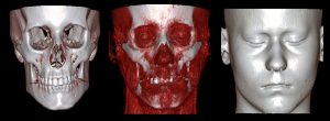
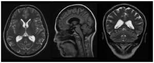
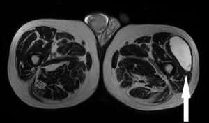
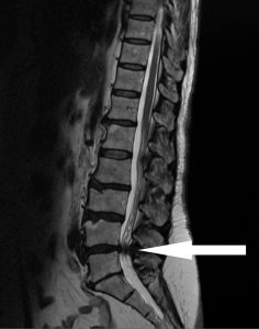
- Section MRI - Magnetic Resonance Imaging
Characteristics and principle of the method
Magnetic resonance imaging (MRI) is one of the modern imaging methods. It is distinguished by its excellent contrast resolution of individual tissues, i.e. the ability to distinguish even tissues with very similar structures. In practice, this means not only, for example, excellent differentiation of the white and grey matter of the brain, but (more importantly) differentiation of normal tissue from tissue affected by a disease process. In this respect, magnetic resonance imaging has a privileged position among all imaging methods. Translated with DeepL.com (free version)
The principle of MRI is different from other imaging methods. It uses the specific physical properties of the nuclei of hydrogen atoms. Hydrogen nuclei, when exposed to a strong magnetic field, are the source of radiofrequency waves. These waves (among other things, very similar to the waves used to transmit radio signals in the VHF band) are picked up by a system of receiving coils (antennas).
It is important to mention that no harmful side effects of magnetic resonance imaging on the human body have been proven so far, it is possible from the 4th month of pregnancy!
The price of 1 examination in the Czech Republic is currently around 7 000 CZK.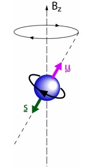
Examination procedure
The examination begins with placing the patient on the examination table. The instruments are divided into closed and open. In the former, the patient is placed in a relatively confined space, which can be uncomfortable, especially for claustrophobic patients. The advantage, however, is the possibility of taking better quality images in a shorter time, as these devices have a stronger magnetic field.
During the examination, a contrast agent may be injected into a vein. Contrast agents used for MRI most often contain gadolinium compounds, rarely manganese or iron. The risk of an allergic reaction to these substances is extremely low. MRI does not use contrast agents containing iodine.
The duration of the MRI scan is in most cases between 20 and 50 minutes.Preparation for examination
Preparation is practically not necessary. The patient does not need to be hungry before the intravenous administration of the contrast agent. Claustrophobic patients are advised to come to the examination with a companion (even a strong feeling of claustrophobia can be easily removed by applying a light sedative, but you must not drive or perform activities requiring concentration after the application).
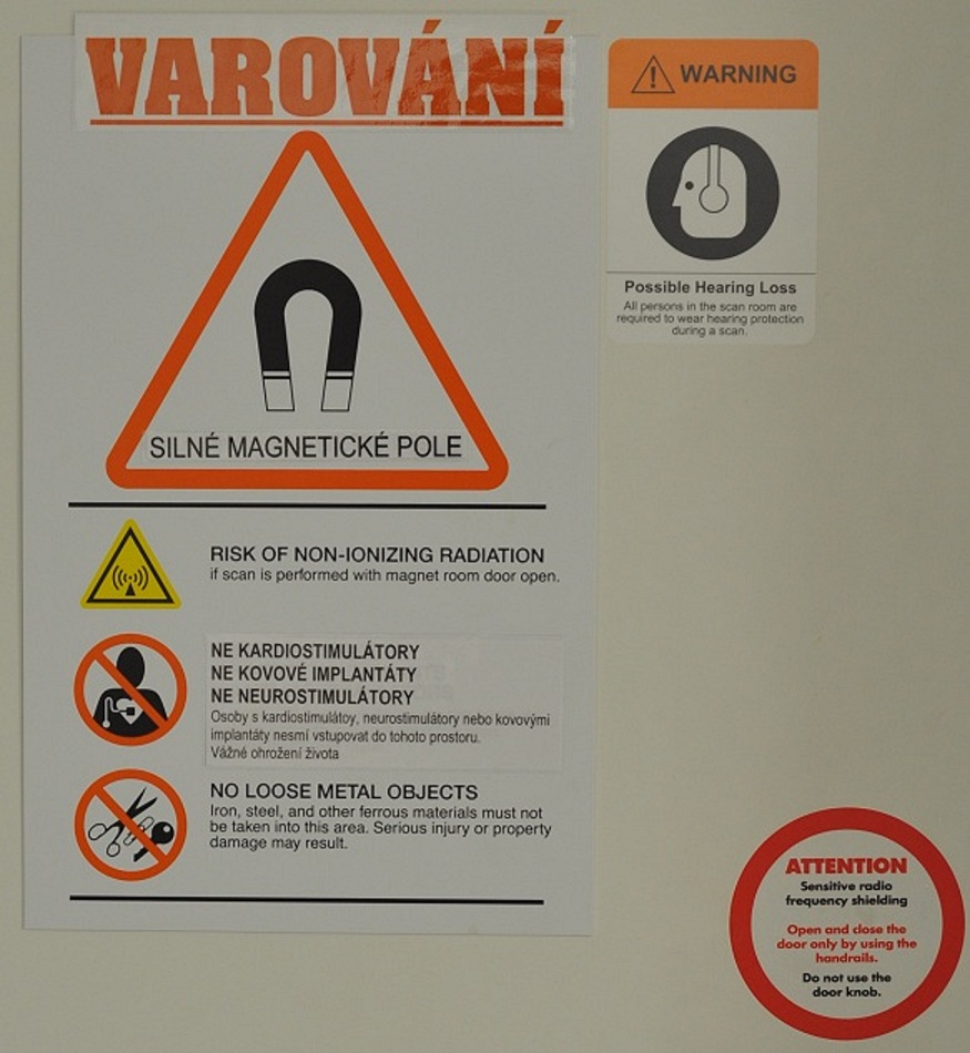
If you are wearing any electronic or metallic implant or foreign body, it does not automatically mean that you cannot have an MRI scan. However, you must ALWAYS and BEFORE the examination report this fact to the operator of the MRI machine who will make an informed decision as to whether or not you can have the examination.Use of the method (indications)
The use of MR is very wide, from classical imaging of the central nervous system to imaging of blood vessels (MR angiography), joints, organs of the chest (heart) and abdomen or other special techniques such as MR spectroscopy, functional MR of the brain, MR diffusion imaging, etc.
When the examination is not appropriate (contraindications)
MRI scans cannot be performed on people with certain types of electronic or metal implants or foreign bodies. In particular, patients with a pacemaker or implanted cardioverter defibrillator (ICD) cannot be examined because there is a risk of serious impairment of its function or serious or even life-threatening disturbance of the heart rhythm. Persons with metallic vascular clamps after cerebral artery aneurysm (aneurysm) surgery can be examined by MRI only under strictly specified conditions.
Where the examination is performed
Examinations are performed by specialized accredited radiological workplaces, our workplace is accredited by the Radiological Society of ČLS JEP, accredited by the SAK of the Czech Republic and ISO 9001:2001 quality certificate within the whole Hospital Jihlava.
You can make an appointment / inquire about an examination by calling 567 157 281
Instrumentation and sample images:
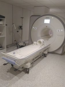
Department of Imaging Methods